Deze samenvatting
+380.000 andere samenvattingen
Een unieke studietool
Een oefentool voor deze samenvatting
Studiecoaching met filmpjes
Onthoud sneller, leer beter. Wetenschappelijk bewezen.

Samenvatting - Class notes - Nutritional Neurosciences
-
1609714800 L1+L2
-
- Food is necessary for survival
- Food is a primary reward
- Food digestion/absorption is a homeostatic challenge!
- A primary brain function is maintaining homeostasis
- The brain strives to automate responses & minimise cognitive 'interference'
- Food is necessary for survival
-
You eat with your brain
-
Where is the human nervous system build off?
- Central nervous system
- Brain
- Spinal cord
- Peripheral nervous system
- Peripheral nerve
- Central nervous system
-
Name the 8 blood vessels in the brain
- Common carotid
- External carotid
- Internal carotid
- Vertebral
- Basilar
- Posterior cerebral
- Middel cerebral
- Anterior cerebral
- Common carotid
-
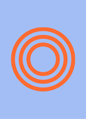
Besides the 8 blood vessels in the brain you also have the circle of Willis, what is this?It is a kind of roundabout. All the blood goes via this...? If there is a blockade somewhere the circle of willis can still provide blood to go to the region.
green part in the picture ...? -
Blood brings 3 things to the brain:
- Glucose
- Oxygen
- Hormones /
signalling molecules
(antibodies/co2) - Glucose
-
The brain exists of three things
- Forebrain
- Brainstem
- Cerebellum
- Forebrain
-
The brainstem consists of:
- Midbrain
- Pons
- Medulla
- Midbrain
-
What are the brain stem functions?
- Reward processing
- Processing gut signals
- Control of heart and breathing rate
(Motor control NIET) - Reward processing
-
Brainstem
- Receives/processes multiple GI signals
- Midbrain:
- Autonomic functions
- Substantia nigra & VTA contain dopamine neurons ->reward circuit
-
What are the functions of the Cerebellum (small brain)
- Motor control:
classis - well established- Cognitive functions:
- Mounting evidence (=steeds meer
bewijs ) - Feeding control:
- Mounting evidence (=steeds meer
bewijs )
May link somatic and visceral systems. Under investigation.... -
The forebrain consists of (is divided in):
- Cerebral cortex
- Basal ganglia
Diencephalon
- Cerebral cortex
-
One part of the forebrain is the cerebral cortex. The cerebral cortex is divided in:Two hemispheres
- 4-5 lobes
- Frontal lobe
- Parietal lobe
- Temporal lobe
- Occipital lobe
- Limbic 'lobe' / limbic system
- connected by the corpus callosum
-
Why is the white matter white?Because of myelin
White inside, gay outside -
One part of the forebrain is diencephalon, in which two parts is diencephalon divided?
- Thalamus
- Hypothalamus
- Thalamus
-
What are the terms to navigate the brain?
- Medial: near the midline
- Middle
- Lateral: near the outer edge
- Dorsal = superior
- Ventral = interior
- Medial: near the midline
-
Where is this blob located?Ventromedial
-
There are three ways to slice the brain
- Transverse = axial
- Coronal
- Sagittal
- Transverse = axial
-
Two ways to view the brain
- Radiological (R=L): upward from the feet
- Neurological (R=R): downward from the head ("brain surgeon view")
- Radiological (R=L): upward from the feet
-
Hoe kan je er zeker van zijn dat je weet welke kant links en welke kant rechts is van het breinDoor het toevoegen van een marker
-
In what kind of ways is there brain nomenclature options
- Brodmann areas
- Anatomical label
- Anatomical location
- Functional name
- (Cytoarchitectonic name)
- Brodmann areas
-
Brain atlases - 'standard space'
- Common reference
- Needed for 'pooling' brains in group analyses
- 'normalization' = warping
- Common reference
-
Talairach space
- Founder: Jean
Talairach - Based on the brain of one 60-y old French woman
Talairach &Tournoux atlases
3D ; stereotaxic coordinate system- Origin at Anterior
Commissure (=midsagittal), Ac-PC line is the y-axis, should be horizontal
Nadeel: gebaseerd op 1 brein - Founder: Jean
-
MNI space
- Montreal
Neurological Institute space: MNI space - Roughly equivalent to Talairach space, same origin
- Brain templates based on average anatomical MRI's of healthy right-handed volunteers acquired by the International Consortium for Brain Mapping (ICBM; n=305 / 152 / 452)
- Montreal
-
What are the notes on
atlases - Very useful, but:
- There is great inter individual aeration in
Sulci andgerij : every cortex is unique- Size of ventricles
- Affected by many factors
- -->
Group-specific (MNI ) templates - --> Probability atlases
-
Gender differences in brain morphology and function can be substantial!
-
What are the gender differences
- Greatest effects in regions affected by sex hormones during brain development
- Several fMRI studies show effect of menstrual cycle/sex hormones on food reward responses
- Greatest effects in regions affected by sex hormones during brain development
-
Diet has also effect on the brain
-
What is the biggest
lobe ? And wat are the functions of thislobe ?Frontallobe Stimulus evaluation, decision making- Controlling movement - planning behaviour
-
What are the relevant subparts of the frontal lobe?
- Orbitofrontal cortex
- Dorsolateral prefrontal cortex
- Medial prefrontal cortex
- Orbitofrontal cortex
-
Which
lobe is next to the frontallobe ? And what are its functionsParietal lobeSomatosensory processing- Controls 'bodily sensations'
- Visual processing (dorsal stream = 'where')
-
Where is the temporal lobe located? And what are its functions
- Auditory processing
- Visual processing (ventral stream = 'what')
- Hippocampus (limbic area)
- Auditory processing
-
Where is the occipital lobe located? And what are its function(s)Visual processing
-
Where is the
insula located?- Insular cortex (
insula = island) - Hidden in the lateral
sulcus - Concealed by parts of frontal, parietal & temporal lobes
Operculum = 'lid' e.g. Frontal operculum
- Insular cortex (
-
What are the functions of the insula
- '
limbis ' area, emotion, interception, homeostasis - anterior
insula : olfactory,gustatory and limbic function; subjective feelings - posterior
insula : perception of bodily sensations, e.g. Pain, visceral sensations, gastricdistension - key connecting area. Connects with
S2 , amygdala, prefrontal cortex, ...
- '
-
What is the '5th lobe' ? And what are its functionsLimbic system
- Subcortical
- Emotion, learning, motivation, autonomic functions
- Key players: amygdala, hippocampus, hypothalamus
- other/associated areas include
- Basal ganglia ('reward')
- Orbitofrontal cortex
- Piriform cortex (smell)
-
Which limbic brain structure is this?Hippocampus
-
What are the functions of the hippocampus and there is it located
- Hippocampus = 'sea horse'
- Location: medial temporal lobe
- Formation of memories
- Forming associations; (dietary) learning
- Spatial navigation
-
What are the functions of amygdala and where are they located?
- 'almond'
- controls autonomic, emotional and sexual behaviour
- Fear, arousal - emotion (unpleasant as well as pleasant)
- Couples learned cues to adaptive response
- Valence (relevance); e.g. Hunger modulates Am response to food stimuli
-
What is the location of the basal ganglia and what are its functions
- Motor control!
- Reward processing - approach/avoidance
- Striatum = putamen +
pallidum +caudate - Dorsal striatum
- Ventral striatum
- nucleus
accumens --> 'hedonic hotspot'
- Motor control!
-
Where is the Thalamus locatedSensory relay
-
Where is the hypothalamus located and what are its functions?
- Homeastasis!
- Integration with hormones
- Energy intake regulation
- Hunger
- Thirst
- Much more; stress, slee, ...
- Homeastasis!
-
5 characteristics of the hypothalamus
- Integration of neural and hormonal signals
- Sensitive to glucose
- Sensitive to blood-borne hormones e.g.: insulin, leptin, gherkin, PYY, CCK, GLP-1
- Multiple neuropeptides e.g.: NPY, AgRP/POMC, alfaMSH
- Implicated in energy intake regulation; hunger (lat), satiety (vm)
- Integration of neural and hormonal signals
-
Spinal cord consists of ...
- Cranial nerves - ascending and descending white matter tracts
- (dorsal horn) - sensory and interneurons
- (ventral horn) - motor neurons that project to muscles
- Cranial nerves - ascending and descending white matter tracts
-
How many cranial nerves are there?
- 12 cranial nerves
- Receptor cells --> sensors, 'senses'
- Autonomic & somatic part
- Olfactory
- optic
- oculomotor
- trochlear
- trigeminal
- abducens
- facial
- auditory
- glossopharyngeal
- vagus
- spinal accessory
- hypoglossal
- 12 cranial nerves
-
Where does the autonomic nervous system contains of?
- Parasympathetic
- Rest & digest/maintenance
- Acetylcholine
- Sympathetic
- Action (fight/flight/freeze)
- Norepinephrine (=noradrenaline)
- Enteric nervous system
- Parasympathetic
-
Peripheral nervous sytem -->
- Sensory perception, nutrient sensing --> cephalic phase responses
- Most relevant nerves: 'sensory' nerves in the head and the vagus nerve (X)
- Sensory perception, nutrient sensing --> cephalic phase responses
-
What is needed for vision?
- Cranial nerve II
- Thalamic nuclei
- Primary & secondary visual cortex in occipital & temporal/parietal lobe
- Visual processing e.g. Attention is modulated by frontal cortex; top-down control
- Cranial nerve II
-
Which regions will get active when looking at food?
- Visual cortex
- Posterior insula
- Amygdala
- Orbitofrontal cortex
Modulators:- Hunger
- BMI
- Attention
- Visual cortex
-
How is the olfaction registered?
- Olfactory nerve (CN I)
- Olfactory bulb --> tract
- Piriform cortex
- Orbitofrontal cortex (insula, amygdala, hippocampus, striatum, thalamus, thupthalamus
Geur gaat direct de hersenen in en komt niet via de thalamus eerst - Olfactory nerve (CN I)
Lees volledige samenvatting
Deze samenvatting. +380.000 andere samenvattingen. Een unieke studietool. Een oefentool voor deze samenvatting. Studiecoaching met filmpjes.
Laatst toegevoegde flashcards
What can you do with fMRI?
- Localisation of function --> Where?
- Function/process --> Multiple regions, response pattern
- Changes over time --> Repeated measurements
- Intervention effects --> Food/drug/training effects
- Functional connectivity --> Network analysis (tast or resting state)
What are the con's of fMRI?
- BOLD signal is an indirect (vascular) measure of neuronal activity
- BOLD response (hrs) is slow; low temporal resolution
- Task design is crucial and far from 'naturalistic'
- Scan environment + magnetic field pose restrictions
- Relatively expensive + time consuming
Nevertheless, very popular
What are the pro's of fMRI?
- Non-invasive, accessible method to study the 'brain in action'
- Repeated measures possible
- Can reveal adaptive reorganisation/changes in the brain, not (yet) detectable in behaviour
- Can reveal functional connectivity between brain regions
Where stands BOLD fMRI for
Blood Oxygenation Level-Dependent (BOLD) signal (indirect measure of (change in) neuronal activity)
^ neural activity --> ^ blood oxygen --> ^ fMRI signal
What is the difference between MRI and functional MRI (fMRI)
MRI studies brain anatomy
Functional MRI (fMRI ) studies brain function
BOLD fMRI = Blood oxygenation Level-Dependent (BOLD) signal (indirect measure of (change in) neuronal activity)
Functional MRI (
BOLD fMRI = Blood oxygenation Level-Dependent (BOLD) signal (indirect measure of (change in) neuronal activity)
Image acquisition =
T1 and T2 are tissue-specific 'relaxation times' of the MRI signal and differences in T1/T2 are used to generate different contrast
Same anatomy, different contrast. Due to difference in T1/T2 relaxation between fat and water
The anatomy of an MRI scan
- Place object in strong static magnetic field, then:
- Transmit RF pulse; excitation
- Wait
- 'Listen' to PF emission due to relaxation (receive)
- Wait, then go to 1 again
- time between step 1 and 3 is Echo Time (TE)
- Wait time before step 1 is repeated: Repetition Time (TR)
- TE and TR influence image contrast differently
Slice selection gradient =
- Apply a gradient along the z-axis
- Larmor frequency differs along the gradient; stronger B0, higher Larmor frequency
- RF pulse only energies the part where field strength matches Larmor frequency (=slice)
- 'Frequency encoding'
Which 3 gradient coils can be made?
z, x and y
Gradient =
Gradients
- add a gradient pulse to code another direction
- 'phase encoding' - changes phase of the spins
Gradient switching - that's what makes all the noise during imaging